Next: Conclusions Up: Independent Component Analysis For Previous: An Inverse EEG Problem
Numerical Simulations
We prepared the simulated data as described in the previous sections. The time-dependent course of 180 ms for all 32 channels is shown in Fig. 6. We also provide a color mapped plot of the potentials on the surface of the head for the time step at 160 ms (maximum variance) in Fig. 12. As can be seen in the latter figure, the distribution of potentials on the scalp can hardly be attributed to a single dipole, but rather to a configuration of several dipoles. We perform PCA on the original EEG time-dependent data and the singular values are shown in Fig.7. Analyzing the singular values, we can deduce that the signal subspace consists of the four first singular vectors. Working with just the contribution of these four components Eq. (9), we perform the ICA procedure, resulting in the activation maps shown in Fig. 8. Notice that there are only three different activation patterns presented; the fourth component is actually just noise.Projecting the first activation back onto all 32 channels, we get the signals shown in Fig. 9, which are the potentials due to the single temporal lobe dipole. Plotting the potentials again for the time step at 160 ms in Fig. 13, one can recognize the surface potential map as resulting from the activation of a single dipole source. This is evidenced by the well-defined foci near the right eye and ear, as well as the symmetric potential fall-off about the dipole plane.
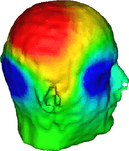
Figure: Scalp surface potential map due to several dipoles,
corresponding to time T=160 ms from the signals shown in Fig. 6.
We can check the accuracy of the ICA decomposition by comparing the
above results to the results of the forward simulation run with the two
other dipoles ``turned off''. Because ICA does not preserve scale, we use
time-space correlation coefficients as our metric for comparing the potentials
at the electrodes. The sets of electrode potentials are viewed as vectors
in time-space and the cosine of the ``angle'' between them is calculated
by taking the dot-product of the two vectors after they have been normalized.
Evaluated this way, our three activation projections restored the original
(unmixed) potential distribution with RMS errors of ![]() ,
, ![]() and
and ![]() .
.
We now turn our attention to the last step of the procedure: source localization. For our head model, on average, the downhill simplex algorithm required only 2-3 interactive restarts in order to converge to the correct solution, with an average run of 30-50 iterations. This is a substantial speed-up compared to the batch-mode multi-start multiple dipole localization methods reported in [36]. The localized temporal lobe dipole was found to be accurate within 4 mm of the actual source. We repeated this localization procedure for the occipital lobe and Sylvian fissure dipoles and were able to determine their positions with errors of 5 and 2 mm, respectively.

Figure: Projection of the first ICA component onto the 32 channels
at the time T = 160 ms.
Next: Conclusions Up: Independent Component Analysis For Previous: An Inverse EEG Problem Zhukov Leonid
Fri Oct 8 13:55:47 MDT 1999