Next: Conclusions Up: Heart-Muscle Fiber Reconstruction from Previous: Rendering

Discussion

The mammalian heart primarily consists of four chambers: two ventricles at the bottom of the heart, and two atria above [Streeter 1979]. The heart also contains other structures, such as the valves between the chambers and the papillary muscles controlling the valves. The heart pumps the blood along a multi-part pathway between the atria and ventricles. Since a muscle fiber can contract only in one direction, the heart structure is complex, to succeed at pumping the blood. Anatomically, to achieve this, the muscle walls of the ventricles and atria are composed of a single helically folded muscular structure, whose fibers continuously connect from one part of the heart to another [Streeter et al. 1978]. A contraction wave passes through this muscular helix, which creates the complex pumping cycle of the heart. The details in the geometric aspects of the helical structure determines the proper functioning of the heart. It is proposed that a better understanding of this helical structure can be used to more effectively correct impaired heart function with surgery [Buckberg et al. 2001]. Physically, muscle consists of many honeycombed ``sarcomeres,'' consisting of interdigitating myosin filaments, in an actin matrix. Water molecules have difficulty in crossing cell membranes and the sarcomere boundaries. It is much easier for water molecules to diffuse to different parts within the same cell and within the same sarcomere than it is for them to cross into other cells or into other sarcomeres. Thus, in muscle tissue, water molecules diffuse in a preferential direction - they are more free to move up and down the length of the muscle fiber. Due to these diffusion properties, muscle tissue can be characterized by one somewhat larger eigenvalue of the diffusion tensor, whose eigenvector is associated with the axial direction of the fiber, and two somewhat smaller eigenvalues; the eigenvectors for these should be perpendicular to the direction of the muscle fiber. Since there is a great deal of structure within the sarcomere, the large eigenvalue and the smaller eigenvalues are not vastly different from one another. Our main results are presented in Figs. (4-6). Heart muscle fibers in Fig. (4) are colored according to RGB-XYZ color scheme. The spiraling diagonal orientation of the muscle fibers on the inside and outside surfaces of the heart reaches $\sim 60$ degrees angle with respect to the vertical. In Figs. (5- 6) the color scheme is sensitive to clock/counterclockwise direction of the spiraling muscle fibers. Clockwise spiral orientation on the inside surface of the heart (endocardium) shown in purple , and counterclockwise muscle fiber orientation on the outside surface of the heart (epicardium) is shown in blue.


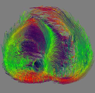
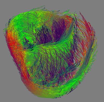
Figure 4: Reconstruction of heart muscle fibers using MLS algorithm. This image uses the RGB-XYZ color scheme. Notice spiraling diagonal orientation of the muscle fibers on the inside and outside surfaces of the heart.


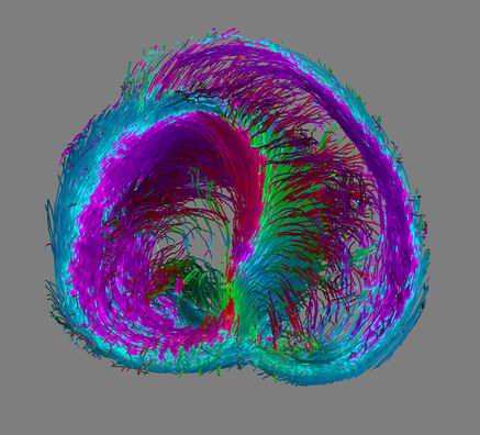
Figure 5: In this figure we use a color scheme which is sensitive to clock/counterclockwise direction of spiraling muscle fibers. Clockwise spiral orientation on the inside surface of the heart (endocardium) shown in purple, and counterclockwise muscle fiber orientation on the outside surface of the heart (epicardium) is shown in blue. This result illustrates the hypothesis that the spiraling fiber orientation in the heart muscle changes from endocardium to epicardium. The fibers are color-mapped independently on how steep the pitch is, causing abrupt change of color on the top.


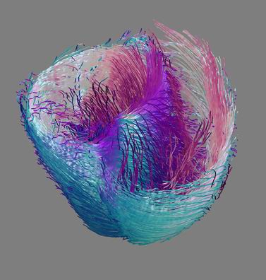
Figure 6: The color scheme used in this figure changes smoothly from clockwise to counterclockwise spiral oriented fibers. Horizontal parts (very small pitch angle) of the fibers are shown in white. This coloration is consistent with observations of some heart researchers, who have described a systematic smooth variation in pitch and direction of heart muscle fibers from endocardium to epicardium.


Next: Conclusions Up: Heart-Muscle Fiber Reconstruction from Previous: Rendering
Leonid Zhukov 2003-09-10