Next: Tensor Classification
Up: Method
Previous: Method
Diffusion tensor magnetic resonance imaging (DT-MRI) [1]
is a technique used to measure the anisotropic diffusion properties of
the water molecules found within biological tissues as a function of
the spatial position within the sample. Due to differing cell shape
and cell membrane properties, the diffusion rates of the water
molecules are different in different directions and locations.
For instance, neural fibers are comprised mostly of bundles of long
cylindrical cells that are filled with fluid and are bounded by
less-water-permeable cell membranes. The average diffusion rate (at a
spatial location) is fastest in the three-dimensional axis direction along
the length of the neuron cells, since more of the water molecules are free
to move in this direction. The average diffusion rate is slowest in the
two transverse directions, where the cell membrane interferes, reducing
and slowing down the movement of the water molecules.
Other parts of the brain are primarily comprised of fluid without cell
membranes, such as the ventricles. Here the average diffusion rate is
larger and more uniform (almost the same in all directions).
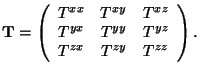
The diffusion properties can be represented with a symmetric second order
tensor -  x matrix:
|
 |
|
|
(1) |
The 6 independent values (the tensor is symmetric) of the tensor
elements vary continuously with spatial location.
The 3-D local axis direction of the neuron fibers will correspond to
the dominant eigenvector of the tensor. There should be one large
eigenvalue, and two small eigenvalues. This can be seen from the
physical interpretation of the diffusion tensor, which can be thought
of as a vector-valued function whose input is the local 3-D
concentration gradient and whose output is the 3-D directional vector
flux2
of the water molecules. The function is
evaluated by multiplying the 3x3 matrix by the 3x1 concentration
gradient, producing the 3x1 vector flux of the water molecules. Water
will diffuse fastest in the direction along the axis of the neurons
and slowest in the two transverse directions.
For the ventricles, a dominant eigenvector should not exist: the three
eigenvalues of the tensor should have roughly the same value. Water will
diffuse roughly at the same speed in all directions. Hence, we can use the
diffusion tensor to distinguish tissues with a primary diffusion axis from
parts that do not.
In this paper, the experimental dataset contains sampled values of the
diffusion tensor on a regularly spaced grid of
 x
x x
x (cubic)
voxels. We will denote these given tensor values as
(cubic)
voxels. We will denote these given tensor values as
 , where
 and
and
 are the three
dimensional tensor components
are the three
dimensional tensor components
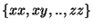 , and
, and
 are
traditional integer indexes into the regular grid volume. Also, when no
upper indexes are provided, the operations are assumed to be
performed on the entire tensor component-wise
are
traditional integer indexes into the regular grid volume. Also, when no
upper indexes are provided, the operations are assumed to be
performed on the entire tensor component-wise
 , i.e., on each of the six independent values of the tensor.
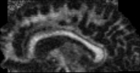
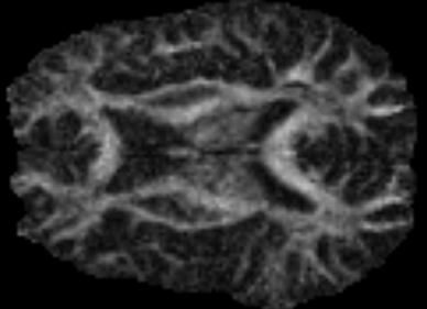
Figure 2:
Sagittal and axial slices of anisotropy measure
 of the
dataset. The lighter regions correspond to stronger anisotropy areas found in the white matter. See Eq.
of the
dataset. The lighter regions correspond to stronger anisotropy areas found in the white matter. See Eq.
 .
.
Next: Tensor Classification
Up: Method
Previous: Method
Leonid Zhukov
2003-01-05